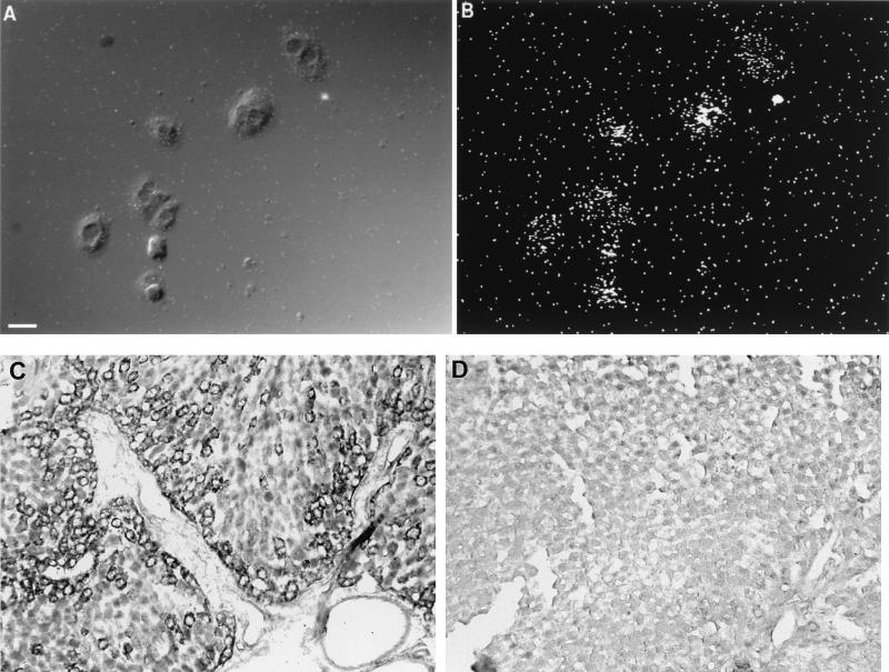
FIG. 3.
Localization of CD14 mRNA in rat primary hepatocytes and sectioned liver. Hepatocytes grown on coverslips or liver tissue sections were subjected to in situ hybridization with antisense and sense riboprobes for CD14. (A) A differential interference contrast image of individual rat primary hepatocytes (the size bar indicates 20 μm). (B) Dark-field view of the in situ signal (35S). Primary hepatocytes isolated from LPS-treated rats (10 mg/kg, i.p., 24-h treatment) show strong cytoplasmic labeling. In the lower panels, in situ hybridizations performed on sectioned livers from LPS-treated (C) and normal (D) rats are shown. The signal detected with the digoxigenin-labeled riboprobe and colorimetric technique shows labeling in individual hepatocytes around blood vessels, in this case a portal triad. (Positive cells on the original slides were blue and, as shown here, are black.) The number of positive cells decreases with distance from the vasculature. In the control liver (panel D), very few or no positively labeled cells are seen. The sense probe showed little or no labeling (data not shown).

