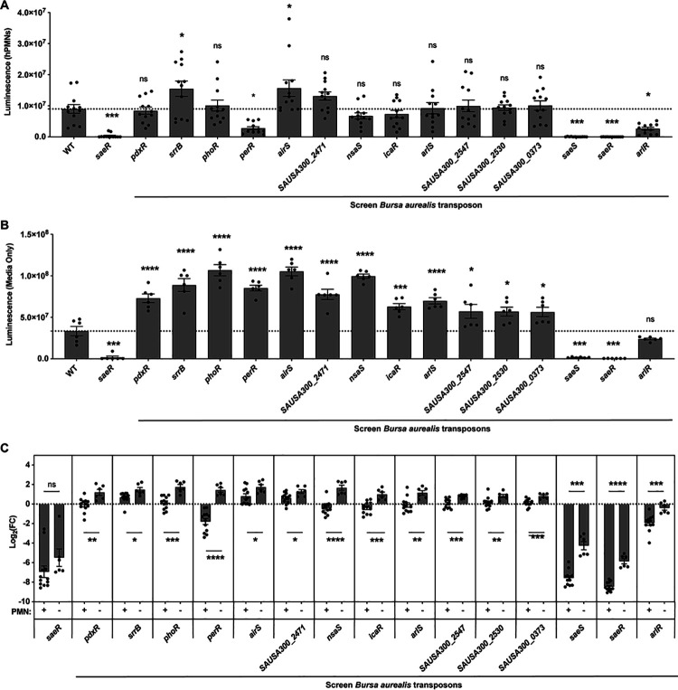
Fig 2.
Regulation of lukAB promoter activity in the presence or absence of hPMNs. (A) PlukAB luminescence values of selected PlukAB activators in the presence of hPMNs in media containing RPMI + HEPES + 5% NHS. The results shown are from two independent experiments each performed with three colonies of each strain repeated in four blood donors (n = 12, MOI = 8). The dotted line represents wild-type JE2. Statistical analysis was performed using one-way ANOVA with multiple comparisons to determine the statistical significance of mutants compared to wild-type JE2. Error bars indicate SEM. (B) Luminescence values of selected PlukAB activators grown as in panel (A) but in the absence of hPMNs. The results shown are from two independent experiments each performed with three colonies of each strain (n = 6). The dotted line represents wild-type JE2. Statistical analysis was performed using one-way ANOVA with multiple comparisons to determine the statistical significance of mutants compared to wild-type JE2. Error bars indicate SEM. (C) Log2 fold change of luminescence of mutants compared to wild-type JE2 in the presence or absence of hPMNs (n = 6–12). Statistical analysis was performed using unpaired t-tests with Welch’s correction to compare the log2 fold change of luminescence in the two conditions for each mutant strain. Log2 fold change of luminescence was used to compare the two conditions to account for the reduction in raw luminescence values because of phagocytosed bacteria, which reduces the efficacy of D-luciferin to cross the bacterial membrane. *P ≤ 0.05; **P ≤ 0.01; ***P ≤ 0.001; ****P ≤ 0.0001. ns, not significant.

