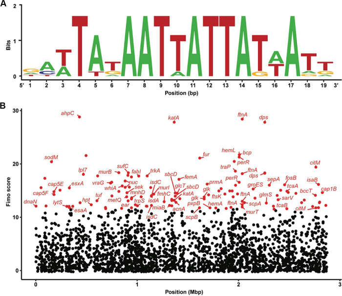
Fig 5.

PerR binding site predicted in many potential genes. (A) PerR binding site sequence motif based on binding site sequences from strain S. aureus 8325-4. (B) Genes predicted to have a PerR binding site. Each dot represents a predicted binding site that lies within a gene or at most 100 bp upstream of it. The horizontal axis represents the coordinates in the assembly at which the binding site occurs, while the vertical axis represents the alignment score; that is how closely the predicted binding site resembles the sequence motif in (A). Genes in red have a FIMO alignment score in the upper 5% and are labeled with their symbol, unless undefined in the reference assembly.
