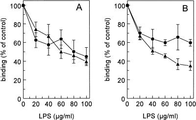
FIG. 5.
Displacement of 125I-oxLDL and 125I-acLDL by LPS. Liver endothelial cells (A) and Kupffer cells (B) were isolated as described in Materials and Methods. Cells were incubated in DMEM–2% BSA (pH 7.4) for 2 h at 4°C with 5 μg of 125I-acLDL (•) or 125I-oxLDL (▴) per ml and increasing amounts of LPS. Cells were then washed, and radioactivity was counted. Data are means of three experiments ± SD.

