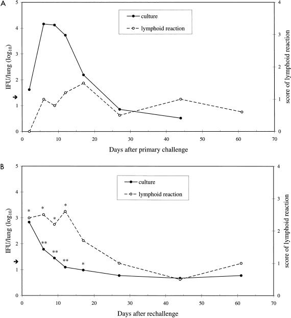
FIG. 1.
C. pneumoniae was cultured from the supernatants of homogenized lung samples obtained at several time points after intranasal primary challenge (A) and rechallenge (B) (given 4 to 8 weeks after primary challenge) of 106 IFU per mouse. The means were calculated from logarithmic data from four individual primary infection experiments and three individual reinfection experiments, with 4 to 10 mice per time point in each experiment. The arrow shows the detection limit for culture (1.3 IFU/lung). The magnitude of the pulmonary lymphoid reaction, evaluated by light microscopy from formalin-fixed lungs from one set of primary infection and reinfection experiments with four to five mice per time point, was scored as 0 (minimal), 1 (mild), 2 (moderate), 3 (marked), or 4 (severe). The asterisks in panel B indicate values that differ significantly between primary infection and reinfection (∗, P < 0.01; ∗∗, P < 0.001).

