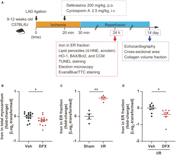
Figure 4. Treatment with deferasirox (DFX) suppresses total myocardial and endoplasmic reticulum (ER) fraction iron contents in I/R‐injured myocardium.

A, Experimental protocol for inducing ischemia reperfusion (I/R) in mice. B, Measurement of total myocardial iron levels 24 hours after DFX administration (200 mg/kg, per os [p.o.]; n=15, each group). C, Measurement of iron levels in the ER fraction of I/R‐injured myocardium 24 hours after I/R (n=4, each group). D, Measurement of iron levels in the ER fraction of I/R‐injured myocardium treated with vehicle or DFX (200 mg/kg, per os [p.o.]; n=8 in vehicle group and 7 in DFX group). Data are presented as the mean±SEM. Statistical significance was determined using unpaired t‐test. *P<0.05, **P<0.01. 4‐HNE indicates 4‐hydroxynonenal; CCM, cleaved caspase substrate motif; HO‐1, heme oxygenase‐1; Hx, hypoxia; LAD, left anterior descending artery; Nx, normoxia; TUNEL, terminal deoxynucleotidyl transferase‐mediated dUTP nick end labeling; and Veh, vehicle.
