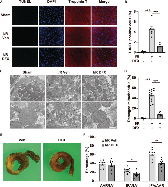
Figure 6. Treatment with deferasirox (DFX) reduces myocardial injury and infarct size in a murine model of ischemia reperfusion (I/R) injury.

A, Representative terminal deoxynucleotidyl transferase‐mediated dUTP nick‐end labeling (TUNEL) staining images of I/R‐injured myocardial tissues from mice treated with vehicle control (Veh) or DFX. Scale bar: 50 μm. B, Quantification of TUNEL‐positive cells 24 hours after reperfusion (n=5 in sham group, n=6 in I/R+Veh, n=6 in I/R+DFX). C, Representative images of I/R‐injured myocardium obtained by electron microscopy. Scale bar: 500 nm. D, Quantification of the number of damaged mitochondria (n=11 in sham group, n=14 in I/R+Veh, n=11 in I/R+DFX). The damaged mitochondria are presented as the proportion of damaged mitochondria to the total number of mitochondria. E, Representative images showing double staining with TTC and Evans Blue in hearts from mice with I/R after treatment with Veh or DFX. Scale bar: 1 mm. F, AAR per LV, IFA per LV, and IFA per L percentages in mice with I/R (n=8, each group). AAR indicates area at risk; IFA, infarct area; and LV, left ventricle. Data are presented as the mean±SEM. Statistical significance was determined using unpaired t‐test. *<0.05, **P<0.01, ***P<0.001.
