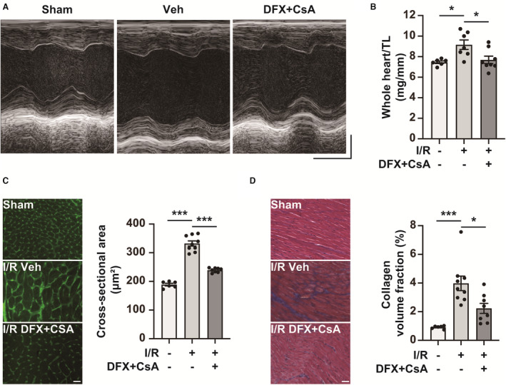
Figure 8. Combination therapy with deferasirox (DFX) and cyclosporin A (CsA) prevents adverse cardiac remodeling in the late phase of ischemia reperfusion (I/R).

A, Representative M‐mode echocardiogram images of sham mice and I/R mice treated with vehicle control (Veh) or DFX+CsA on day 14 after I/R. Horizontal scale bar: 100 msec. Vertical scale bar: 1 mm. B, Whole heart weight per tibial length (TL) of sham mice (n=6) and I/R mice treated with Veh (n=7) or DFX+CsA (n=8) on day 14 after I/R. Two of the I/R mice treated with Veh were excluded because of their death after echocardiography. C, Cross‐sectional area on day 14 after I/R. Representative wheat germ agglutinin staining images of the left ventricle in sham mice and I/R mice treated with Veh or DFX+CsA (left panel). Scale bar: 50 μm. Quantification of cross‐sectional area (right panel; n=6 in sham group, n=9 in Veh group, n=8 in DFX+CsA group). D, Collagen volume fraction (interstitial fibrosis) on day 14 after I/R. Representative Masson trichrome staining of the left ventricle in mice with Veh or DFX+CsA (left panel). Scale bar: 50 μm. Quantification of interstitial fibrosis as the collagen volume fraction (right panel; n=6 in sham group, n=9 in Veh group, n=8 in DFX+CsA group). Data are presented as the mean±SEM. Statistical significance was determined using 1‐way ANOVA with Tukey post hoc test. *P<0.05, ***P<0.001.
