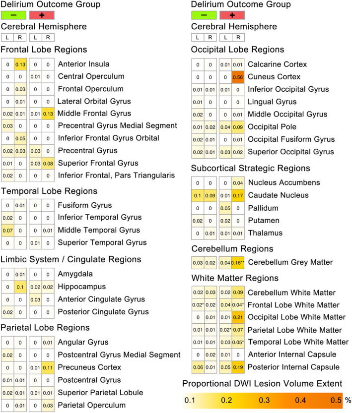
Figure 4.

Regional perioperative diffusion‐weighted imaging (DWI) lesion extent by cerebral hemisphere and delirium outcome group. Heatmap of regional perioperative diffusion‐weighted imaging (DWI) lesion volumes corrected for individual ROI volumes (i.e., DWI lesion volume/ROI volume = regional lesion extent), separated by cerebral hemisphere and delirium outcome group. ROIs with lesion extent >0.01% are shown. Asterisks indicate significant change in regional lesion extent from presurgical baseline; *p‐FDR < 0.01, **p‐FDR < 0.001, ***p‐FDR < 0.0001.
