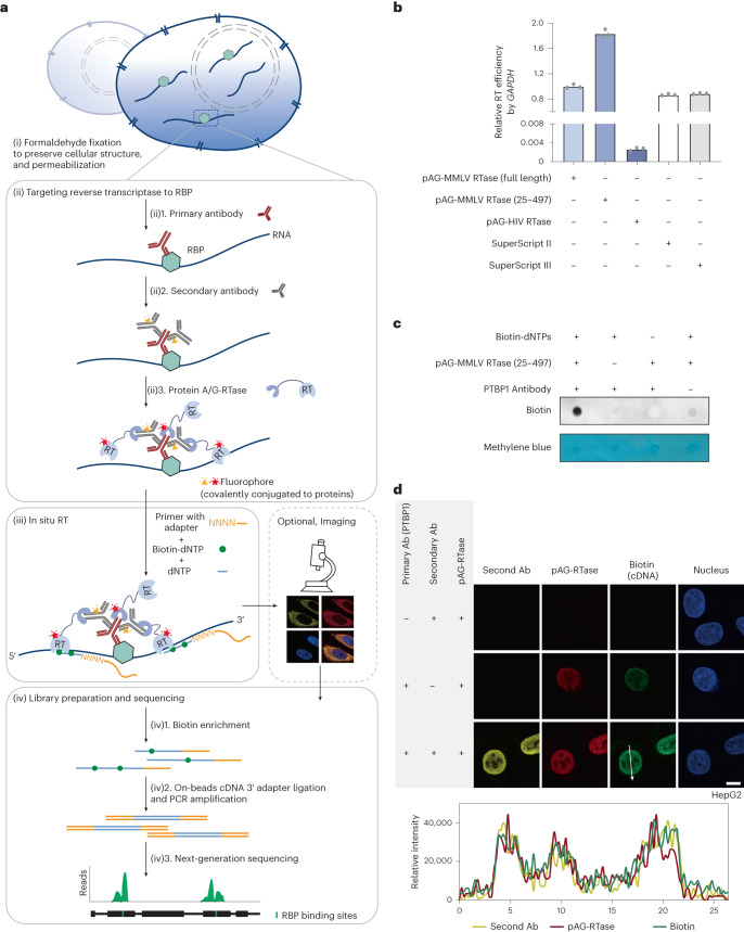
Fig. 1. ARTR-seq strategy and validation.
a, Scheme of ARTR-seq. b, RT–qPCR analysis showing the RT activity of tested purified pAG-RTase fusion proteins. Two commercial RTases, SuperScript II and SuperScript III, were loaded as positive controls. n = 3 biological replicates. c, Biotin dot blot assay showing biotinylated cDNA products produced from ARTR-seq. Methylene blue staining was the loading control. d, Immunofluorescence imaging of the secondary antibody (secondary Ab; yellow), pAG-RTase (red), biotinylated cDNA (green) and nucleus (blue) for PTBP1 ARTR-seq. The line graph analysis shows relative fluorescence intensity along the line. Scale bar, 10 μm.

