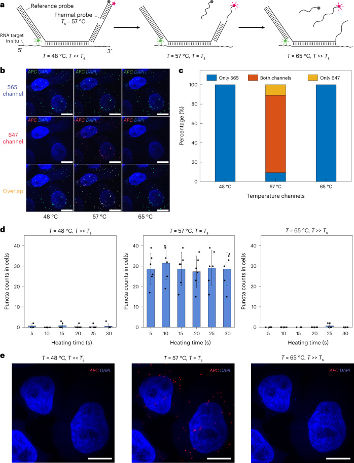
Fig. 2. Validation and fast channel switching speed of DNA thermal-plex for RNA FISH in situ.
a, Design for the reference and thermal probe sets. Reference probes fluoresce at all temperatures, while the thermal probe set fluoresce only after being exposed to a heating spike at its signal temperature (for example, 57 °C). b, FISH imaging of APC RNA transcripts in fixed HeLa cells with a 57 °C thermal probe set in 565-nm channel, 647-nm channel and with both channels overlaid, after transiently exposed to heating spikes at three different temperatures. Only after being heated to the signal temperature 57 °C did the thermal probe show the fluorescence signal, which appear to be colocalized with the reference probe. c, Colocalization analysis of the puncta in 565-nm and 647 channels, showing percentages of the puncta that are detected only in 565-nm channel (blue), only in 647-nm channel (red) and colocalized in both channels (orange). A total of four cells were analyzed. d, Barplots of puncta counts in cells imaged after heating for different amounts of time below (48 °C), at (57 °C) and above (65 °C) the signal temperature. Error bars depict s.d. between cells. e, FISH images of APC gene after heating at different temperatures (48 °C, 57 °C and 65 °C) for 5 s. A total of six cells were analyzed for d. Three independent experiments were repeated for validation. Scale bars, 10 µm.

