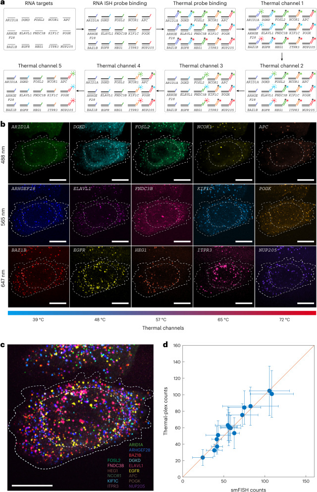
Fig. 4. Fifteen-plex RNA imaging with the combination of five thermal channels and three fluorescence channels in fixed cells.
a, Schematic for 15-plex RNA imaging. Fifteen ISH probe sets with distinct DNA barcodes are hybridized in situ. Fifteen orthogonal DNA thermal probe sets are then hybridized to ISH probe DNA barcodes. Following each iterative round of heating to signal temperatures, fluorescence images in three channels are collected after the sample is cooled to ~30 °C. b, Individual channel images for each of the 15 RNA targets. c, Overlaid images for all 15 mRNAs in a single cell resolved by thermal-plex. See Extended Data Fig. 9 for the computationally reconstructed image. d, Comparison of the resolved 15-plex single-cell RNA expression between thermal-plex and conventional smFISH methods. The red line indicates y = x. The high linear correlation (R2 = 0.95) between the two methods indicates the robustness of the thermal-plex imaging methods. The s.d. shows that the total cell numbers for the thermal-plex and smFISH are 32 and 38 cells, respectively. Data are presented as mean values ± s.d. Scale bars, 10 µm.

