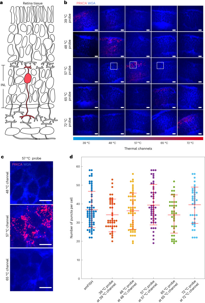
Fig. 5. Thermal-plex imaging in retina tissue.
a, Schematic of retina tissue and targeted cell type (RBCs, red). b, FISH images of the INL of the retinal tissue for five distinct thermal channels at different temperatures (39 °C, 48 °C, 57 °C, 65 °C and 72 °C) all targeting Prkca mRNA. All probes showed strong RNA puncta signal only in their designated temperature channel. The cell membrane was stained with WGA. c, Enlarged images of the retina FISH images with 57 °C thermal probes at three different temperature channels (48 °C, 57 °C and 65 °C). d, Plot of thermal-plex puncta for all five thermal probes in their corresponding thermal channels. smFISH puncta are used as the positive control. All five thermal channels yielded Prkca expression level in the INL region similar to those seen with the conventional smFISH method. The numbers of cells used to count the fluorescent puncta for smFISH and all of five thermal channels were 64, 41, 52, 50, 39 and 40, respectively. Red lines indicate the mean expression level of Prkca mRNA. Data are presented as mean values ± s.d. Scale bars, 10 µm.

