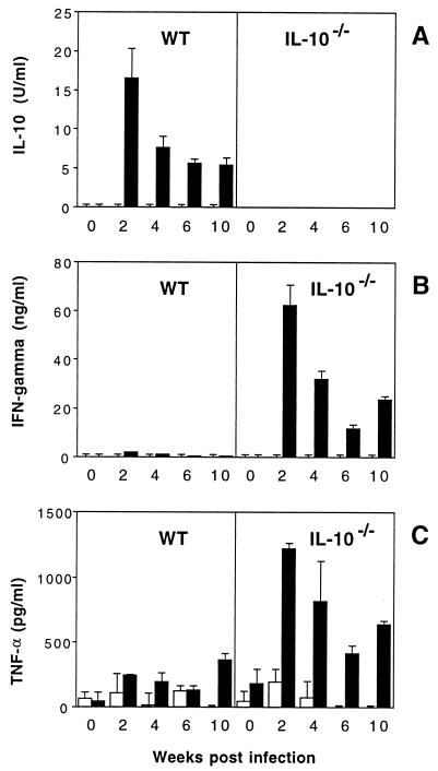
FIG. 6.
SHelAg-induced cytokine responses of MLN cells from H. hepaticus-infected WT and IL-10−/− mice. MLN cells (3 × 10⁶/ml) from uninfected and 2-, 4-, 6-, and 10-week-infected WT and IL-10−/− mice were stimulated with medium alone (□) or 1 μg of SHelAg per ml (■), and IL-10 (A), IFN-γ (B), and TNF-α (C) were measured in 72-h supernatants. Bars represent means ± SD of duplicate ELISA values from a pool of two to three infected WT and three to four infected IL-10−/− mice per time point. Cytokine levels for the 0-week postinfection time point have been calculated after pooling data from three separate determinations for uninfected mice assayed in parallel with the infected mice at 4, 6, and 10 weeks postinfection.

