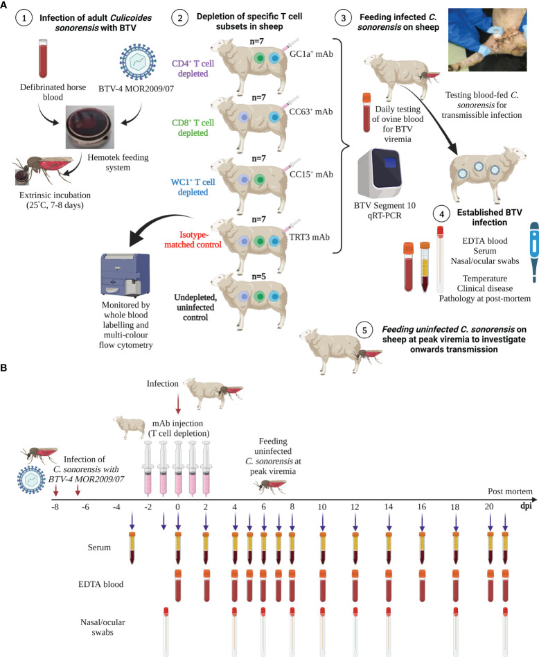
Figure 1.
Schematic diagram of experimental design and timeline. (A) Adult Culicoides sonorensis were fed on a defibrinated horse blood–virus mixture using the Hemotek feeding system to enable uptake of BTV-4 MOR2009/07. C. sonorensis were incubated at 25°C for 7–8 days (extrinsic incubation period) to enable virus dissemination to the salivary gland. Midges were subsequently allowed to blood-feed on sheep depleted of CD4+ (n = 7), CD8+ (n = 7), or WC1+ γδ T cells, which was achieved by administration of specific monoclonal antibodies (mAbs; GC1a, CC63, CC15), or on mock-depleted sheep (n = 7), achieved using an isotype-matched mAb raised against Turkey rhinotracheitis virus (TRT3). One sheep within each experimental replicate (n = 5) was not depleted and not infected (negative control) to monitor for potential contact transmission. Following blood-feeding, engorged C. sonorensis females were collected to determine the numbers of transmissibly infected (TI) individuals that had taken a blood meal by RNA extraction and bluetongue virus (BTV) (segment 10-specific) qRT-PCR. Whole blood was taken daily or every other day for immunophenotyping, using specific T- and B-cell mAbs, to monitor the T-cell depletion and specific immune cell population dynamics during infection. Whole (ethylenediaminetetraacetic acid (EDTA) and heparin) blood, clotted blood (for serum), and nasal and ocular swabs were taken from all sheep at various time points during infection (see panel B) to monitor viraemia dynamics, host antibody responses, and viral shedding. Temperatures and clinical disease scoring were recorded daily. At peak viraemia, uninfected adult C. sonorensis were allowed to blood-feed on each sheep, and engorged females were collected immediately (day 0) to obtain a virus uptake baseline or following extrinsic incubation (day 8) to monitor infection rate and therefore onwards transmission rate to the Culicoides vector. (B) Timeline of BTV infection, T-cell depletion, and sample collection for the representative in vivo BTV transmission study. Schematic images created in BioRender.com.

