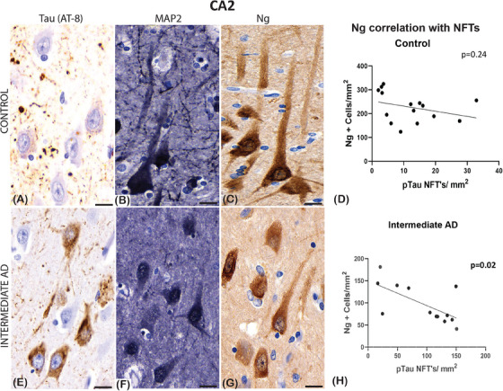
FIGURE 1.

Examples of immunoreactivity of mouse‐anti‐tau (AT‐8), mouse‐anti‐microtubule‐associated protein 2 (MAP2), and mouse‐anti‐neurogranin (Ng) in normal control and in a case with intermediate Alzheimer's disease (AD) in a section from the hippocampal region cornu ammonis (CA2). The control (A) shows sparse apical primary dendritic staining of tau in the pyramidal neurons; in the AD case there are moderate (E) amounts of staining spreading into the apical dendrite. The MAP2 staining was seen in both groups, but the processes are well defined in the control (B) compared to cases of intermediate AD (F). Ng protein expression differs between the groups in pyramidal neurons, as this appears denser and more concentrated around the nucleus of pyramidal neurons in the intermediate AD cases (G), whereas in the control cases the staining throughout the apical dendritic processes (C). No significant Pearson's correlation, in the controls, was seen between the number of Ng2 positive neurons and the number of neurofibrillary tangles (NFTs) defined with phosphorylated tau (pTau; D), whereas a significant negative correlation between Ng and NFTs was seen in the intermediate AD cases (H). Scale bars = 10 μm (A, B, C, E, F, G)
