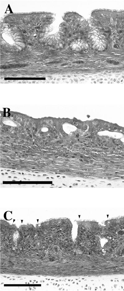
FIG. 1.
Hematoxylin-eosin-stained 4-μm sections of tracheae from 3-week-old turkeys sham inoculated or exposed 2 weeks previously to parent or Dnt− mutants of B. avium, as described in the text. (A) Sham-inoculated control with normal trachea. (B) Parental strain-infected turkey. Trachea shows marked disruption of mucosal architecture; absence of ciliated epithelium and goblet cells; epithelium composed of immature cuboidal cells; glands depleted of mucus; and hyperemia, mild fibrosis, and mixed inflammatory cell infiltrate of lamina propria and submucosa. (C) Dnt− mutant-infected turkey. Trachea shows normal mucosal architecture; normal ciliated epithelium, except for small focal areas of deciliation (arrowheads); partial depletion of mucus from glands; and increased mononuclear cells in lamina propria. Changes are intermediate between sham-inoculated and wild-type-exposed turkeys. Bars, 100 μm.

