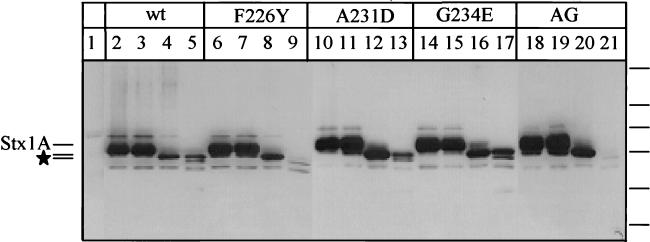
FIG. 2.
Immunoblot of wild-type (wt) or mutant Stx1A following trypsin digestion. Periplasmic extracts of mutant and wild-type Stx1As were incubated at 37°C for 15 min with no trypsin (lanes 2, 6, 10, 14, and 18) or with trypsin at 0.05 (lanes 3, 7, 11, 15, and 19), 0.5 (lanes 4, 8, 12, 16, and 20), or 5 ng/ml (lanes 5, 9, 13, 17, and 21) ng/ml. Lanes: 1, pUC19ΔH (vector-only control); 2 to 5, wild-type Stx1A; 6 to 9, F226Y Stx1A; 10 to 13, A231D Stx1A; 14 to 17, G234E Stx1A; 18 to 21, A231D-G234E Stx1A (AG). The locations of Stx1A and the two primary trypsin degradation products (★) are shown on the left. The positions of the following molecular size standards are indicated on the right (from top to bottom: 95.5, 55, 43, 29, 18.4, and 12.4 kDa).

