
Fig 3.
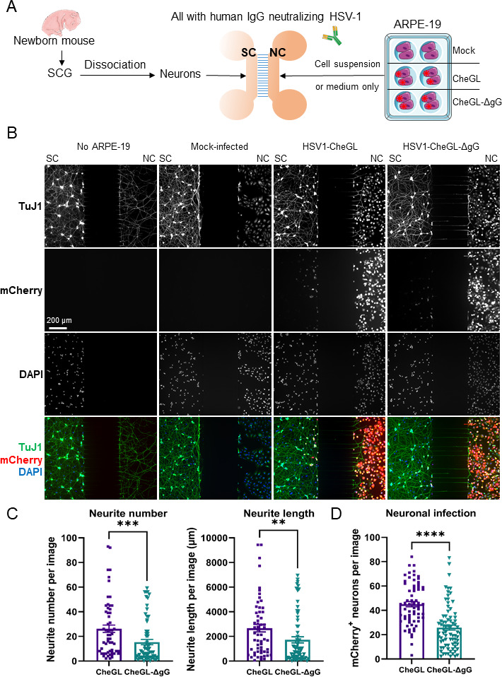
HSV1-CheGL induces more neurite outgrowth than HSV1-CheGL-ΔgG toward infected ARPE-19 cells, facilitating neuronal infection. (A) Schematic representation of the experiment performed to quantify neurite outgrowth and neuronal infection using MFC. SCGs were extracted from neonatal mice and dissociated into single neurons that were seeded into the soma compartment (SC) of MFC. Mock- or HSV-1-infected ARPE-19 cells were added into the neurite compartment (NC) simultaneously. The ARPE-19 cells had been infected with HSV-1 at an MOI of 1 for 16 hours prior to seeding into the NC. The darker color of the NC than the SC indicates that the NC contained more volume to facilitate the diffusion of factors secreted by ARPE-19 cells into the SC. To avoid diffusion of cell-free HSV-1 from infected ARPE-19 cells, we added anti-HSV-1 neutralizing antibodies. (B) Immunofluorescence microscopy images of neurons and ARPE-19 cells grown in MFC. Dissociated SCG neurons were seeded in the SC in all experimental conditions. The absence or presence of mock-, HSV1-CheGL-, or HSV1-CheGL-ΔgG-infected ARPE-19 cells in the NC is indicated above each column. After 20–24 hours, the cells were fixed and labeled with an anti-β-III-tubulin antibody (TuJ1, top row). HSV-1-infected cells were detected by mCherry expression (second row) and nuclei were detected with DAPI staining (third row). These panels show the gray channels, while the bottom row shows images containing all detected signals in their respective colors (TuJ1, green; mCherry, red; DAPI, blue). Scale bar: 200 µm. (C and D) Graphs showing total neurite number and length in the NC of MFCs (C) and number of mCherry positive neurons in the SC (D). Quantification was performed with FIJI software (see Materials and Methods). Each symbol represents the data from one picture taken randomly and covers both sides next to the microgrooves in the MFC. Data are presented as mean ± standard error of the mean. ** P < 0.01, ***P < 0.001, ****P < 0.0001 (Mann-Whitney test). Abbreviations: SCG, superior cervical ganglia; IgG, immunoglobulin; SC, somal compartment; NC, neurite compartment.