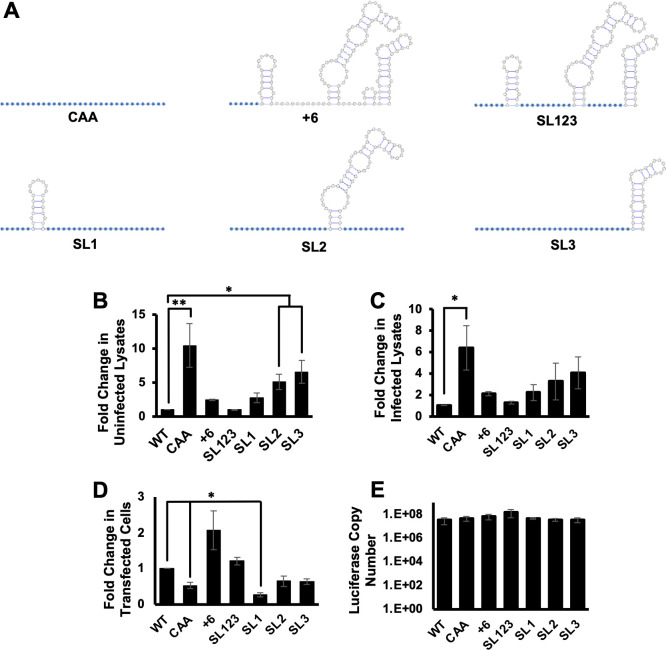
Fig 2.
MIE 5′ UTR secondary structure inhibits translation in vitro. (A) Cartoon showing RNA secondary structure of MIE 5′ UTR mutants. Blue circles indicate nucleotides changed to CAA repeats; white nucleotides are unchanged from WT sequence. (B and C) Caped and polyadenylated in vitro transcribed RNAs containing the WT or mutant MIE 5′ UTR sequences upstream of the luciferase coding region were mixed with cytosolic extracts from uninfected (B) or infected (C) fibroblasts, and the fold change in luciferase activity was measured, with the activity of the reporter containing the WT MIE 5′ UTR set to 1. (D) MIE 5′ UTR luciferase transcripts were transfected into HeLa cells, and luciferase activity was measured. (E) The RNA abundance of each reporter in transfected cells was quantified using RT-qPCR. The graphs show the combined results of three independent experiments (*P < 0.05, **P < 0.01, multiple comparison statistics calculated using Dunnett’s test).

