Fig 1.
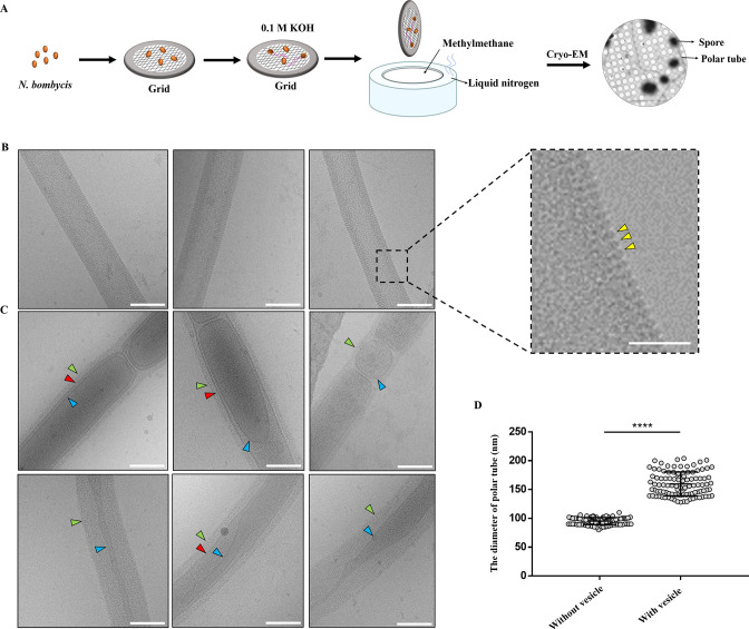
Cryo-EM analysis of the extruded polar tube of N. bombycis. (A) A schematic representation of the methodology for spore germination on the Cryo-EM grid. (B) Cryo-EM analysis of the structural characteristics of the polar tube without vesicles. Bar, 100 nm. The yellow triangles in the enlarged image pointed the bumps distributed on the surface of polar tubes. Bar, 50 nm. (C) Cryo-EM analysis of the structural characteristics of the polar tubes with vesicles. The green triangles pointed the fibrillar material on the surface of polar tubes. The blue triangles pointed the vesicle membrane inside the polar tube. The red triangles pointed the structure of membrane within membrane. Bar, 100 nm. (D) Comparison of the diameter of polar tube with and without vesicle. ****P < 0.0001 (n = 100 for each sample; unpaired Student’s t-test).

