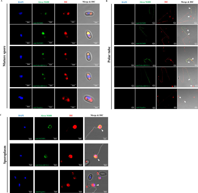
Fig 6.
Immunofluorescence assay of NbTMP1 and NoboABCG1.1 localization in the mature spore, extruded polar tube, and sporoplasm of N. bombycis. (A) Immunofluorescence assay of NbTMP1 and NoboABCG1.1 localization in the mature spore. (B) Immunofluorescence assay of NbTMP1 and NoboABCG1.1 localization in the extruded polar tube. (C) Immunofluorescence assay of NbTMP1 and NoboABCG1.1 localization in the sporoplasm. The mature spore, polar tube, and sporoplasm samples were treated with mouse anti-NbTMP1 serum, mouse anti-NoboABCG1.1 serum, and negative serum, respectively. The nuclei were stained with DAPI (blue), and the membrane structure was stained with DiI (red). The white arrows, diamonds, and triangles indicate the spore empty coat, polar tube, and sporoplasm, respectively. The white dotted line indicates the extruded polar tube. Bar, 2 µm and 5 µm.

