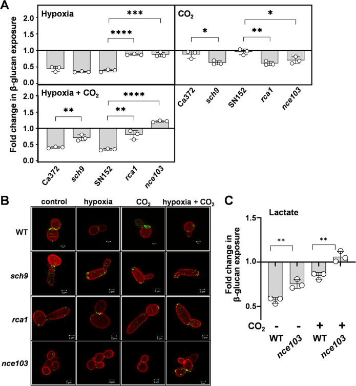
Fig 6.
Impact of Nce103, Rca1, and Sch9 upon the changes in β-1,3-glucan exposure mediated by hypoxia and CO2. C. albicans cells were grown in GYNB at 30°C and exposed to hypoxia, 5% CO2, or a combination of these two inputs. (A) For each strain, the fold changes in β-1,3-glucan exposure were quantified by Fc-dectin-1 staining and flow cytometry, relative to the same strain grown in normoxic GYNB without CO2. C. albicans Ca372 (CAI4 + CIp10) is the wild-type control for sch9 (CAS4), and SN152 is the wild-type control for rca1 (rca1ΔY) (Table S2). Means and standard deviations from three independent replicate experiments are shown, and the data were analyzed using ANOVA with Tukey’s multiple comparison test: *, P < 0.05; **, P < 0.01; ***, P < 0.001; ****, P < 0.0001. (B) Corresponding high-resolution fluorescence confocal images of these cells, double stained with Fc-dectin-1 (exposed β-1,3-glucan, AF488, green) and ConA (mannan, AF647, red). The images are representative of three independent experiments; scale bar = 2 µm. (C) Using an analogous approach, the influence of NCE1 on lactate-induced β-1,3-glucan masking was compared in the presence and absence of 5% CO2 using wild-type (SC5314) and nce103 cells (Table S2). Fold changes in β-1,3-glucan exposure were calculated by dividing the MFI for lactate-exposed cells by the MFI for the corresponding GYNB control (Materials and Methods). Means and standard deviations from three independent replicate experiments are shown, and the data were analyzed using ANOVA with Tukey’s multiple comparison test: **, P < 0.01.

