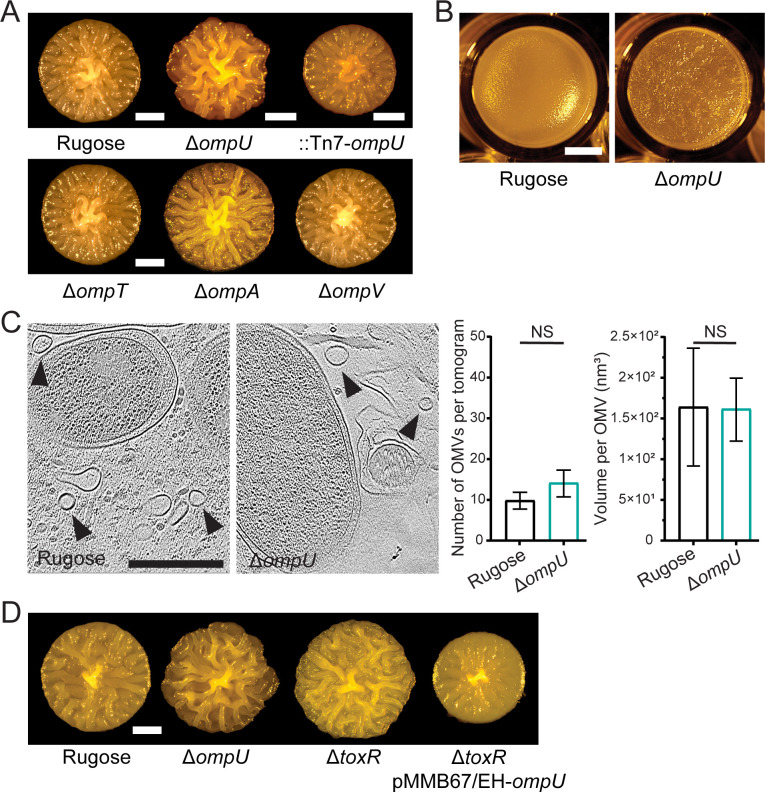
Fig 2.
The outer membrane porin OmpU affects biofilm formation. (A, D) Colony corrugation phenotypes of strains with indicated genotypes after 72 h of growth at 30°C. Images are representative of two biological replicates, with three technical replicates per biological replicate. Scale bars = 1 mm. (B) Morphology of pellicles formed after 24 h incubation at 30°C. Scale bars = 2 mm. (C) Representative cryo-ET tomograms depicting the interior of a biofilm matrix of biofilm grown rugose and ΔompU cells of V. cholerae. Arrows point to the OMVs. Scale bars = 0.5 µm. Average number of OMVs (left) and volume per OMV (right) found in the tomograms (n = 10) of V. cholerae biofilms for the rugose and ΔompU strains, respectively. Error bars represent the standard error of the mean, and statistical significance was determined using Student’s t-test. NS, not significant.

