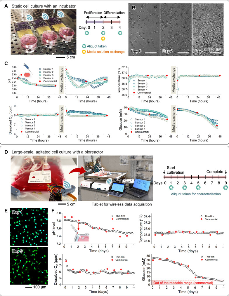
Fig. 6. Demonstration of cell culture with two representative systems.
(A) Static culture of mouse primary myoblasts (mPMs) within a conventional incubator. (B) Representative optical images of mPMs, showing progressive cellular growth (day 2) and subsequent differentiation (day 4) over time. (C) Monitoring results, including pH changes, temperature variations, and levels of the DO and glucose in the cell medium over time. The red-filled circles in all graphs represent the measured values obtained using a commercial sensor or laboratory sensor. (D to F) Large-scale, agitated, GH-encapsulated stem cell culture with a bioreactor. (D) Photo of the flexible sensor-integrated 2-liter cell bag with a rigid prototype of wireless circuit, along with the experimental conditions for hMSC culture in hydrogels within the bioreactor. (E) Representative cell morphology cultured for 9 days in the bioreactor. (F) Monitoring results for a continuous period of 9 days. The red-filled circles in all graphs represent the measured values obtained using a commercial sensor or laboratory sensor.

