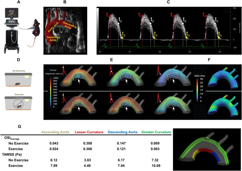
Fig. 2. In silico analysis of exercise-augmented PSS.
Four-dimensional (4D) computational fluid dynamics (CFD) simulation was performed to analyze hemodynamic profiles in the mouse aorta in response to exercise. (A) A schematic of ultrasound system that acquired B-mode and PW Doppler images from the anesthetized mouse. (B) A representative B-mode image and segmentation of the mouse aortic arch and the three branches, and the descending aorta. (C) PW Doppler measurement was acquired from the mouse ascending aorta over four electrocardiogram (ECG)–gated cardiac cycles. (D) Schematics of mice in sedentary condition and engaging in voluntary wheel running. (E) Velocity-colored streamlines along the aortic arch demonstrating disturbed flow developed at the lesser curvature, whereas exercise-augmented shear stress mitigates flow recirculation at the disturbed flow-prone lesser curvature. (F) Time-averaged wall shear stress (TAWSS) over several cardiac cycles was computed and compared between no exercise and voluntary wheel running. Note that PSS develops at the greater curvature of the aortic arch and descending aorta, whereas disturbed flow, including OSS, develops at the lesser curvature. (G) OSIave (averaged over surface) and TAWSS are compared between lesser curvature (ascending aorta, aortic arch, and descending aorta) and greater curvature. Exercise-augmented PSS mitigates disturbed flow developed at the lesser curvature and increases TAWSS in the aortic arch. OSI, oscillatory shear index; Re, Reynolds number.

