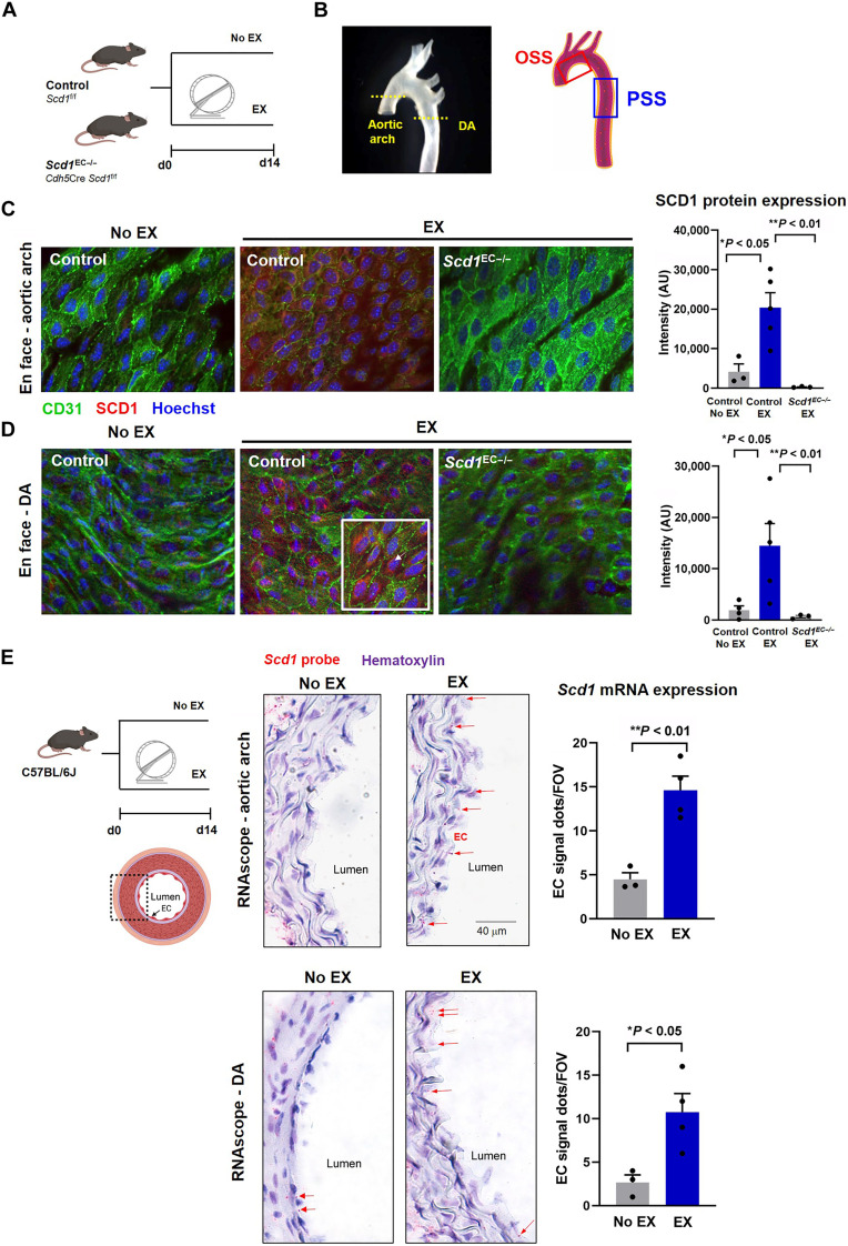
Fig. 4. Exercise activates endothelial SCD1.
(A) Endothelial-specific Scd1-deleted mice underwent 14-day voluntary wheel running protocol. (B) En face immunofluorescence was imaged by confocal microscope at the level of the aortic arch versus descending aorta (DA). (C and D) En face immunostaining of the exposed aortic endothelium reveals that exercise increased SCD1 expression (red) in aortic endothelial cells (green, CD31+) in the aortic arch (C) and the DA (D) (No EX, n = 4; and EX, n = 5; *P < 0.05 versus No EX). At a higher magnification image (×63; bottom right corner inset), SCD1 staining is perinuclearly located (arrows). No SCD1 staining was observed in the Scd1EC−/− mice undergoing exercise in both aortic arch and DA regions. (E) Wild-type C57/BL6J mice underwent 14-day voluntary wheel running protocol. The Scd1-specific probe (RNAscope) in transversal aortic sections demonstrates prominent endothelial staining following 14 days of exercise-induced PSS both in the flow-disturbed aortic arch and the DA. Red dots (arrows) indicate the mRNA molecules in the intima layer. Scd1-positive signals were also present in some smooth muscle cells and periaortic adventitia (adv) in both groups. Microphotographs were taken at ×63 magnification. Quantification is shown on the right panels as the number of positive red dots in the endothelial layer averaged from 5 to 10 microscopic fields per animal (No EX, n = 3; and EX, n = 4; *P < 0.05 and **P < 0.01 versus No EX). FOV, field of view; AU, arbitrary units.

