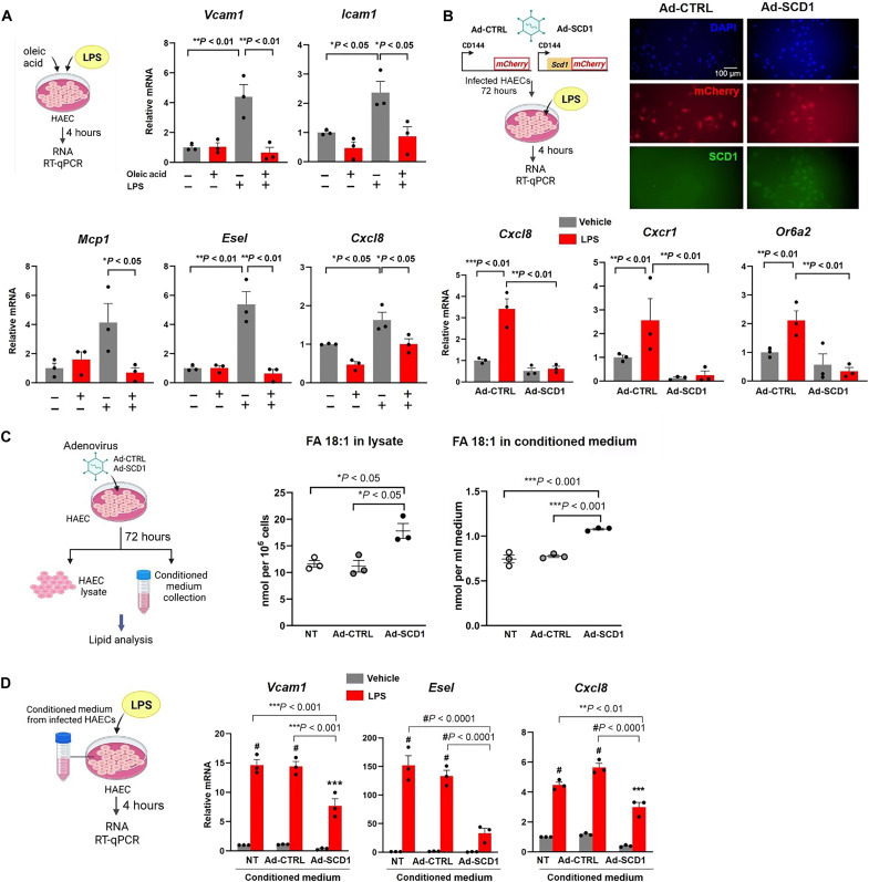
Fig. 5. OA treatment or SCD1 overexpression mitigate pro-inflammatory mediators in HAEC.
(A) HAEC monolayers were treated for 4 hours with OA at 0.2 mM in the absence or presence of lipopolysaccharide (LPS; 20 ng/ml) and reverse transcription quantitative polymerase chain reaction (RT-qPCR) was performed. (B) HAECs were infected with adenovirus control or adenovirus to overexpress SCD1 and subjected to LPS treatment. Transfection was validated using immunostaining for mCherry reporter and SCD1 expression. Ad-SCD1 mitigated the LPS -induced expression (4 hours) of Cxcl8, Cxcr1, and Or6a2 (*P < 0.05, **P < 0.01, and ***P < 0.001; n = 3). DAPI, 4′,6-diamidino-2-phenylindole. (C) HAECs were transfected with adenovirus control or adenovirus to overexpress SCD1. After 72 hours, the conditioned medium, and the cellular lysate were collected and processed for lipidomic analysis. The fatty acid (18:1) content corresponding to OA was normalized to the number of cells in the lysate or the volume of conditioned medium [*P < 0.05, **P < 0.01, and ***P < 0.001; n = 3, one-way analysis of variance (ANOVA)]. Non-transfected cells (NT) were also included as control. (D) HAEC monolayers grown in regular culture medium were changed to conditioned medium collected from HAECs from NT, Ad-CTRL, and Ad-SCD1 in the presence or absence of LPS (20 ng/ml) for 4 hours). RT-qPCR for inflammatory gene expression was performed medium (**P< 0.01, ***P < 0.001, and #P < 0.0001; n = 3, two-way ANOVA).

