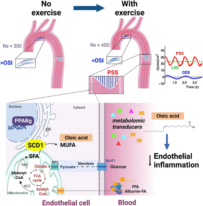
Fig. 9. Schematic model for exercise-augmented PSS in the aorta and activation of the flow-responsive PPARγ-SCD1 pathway for atheroprotective metabolites.
Exercise-mitigates flow recirculation and OSI in the aortic arch. SCD1 located in the ER membrane catalyzes SFA to MUFA, leading to increased intracellular and circulating levels of OA. OA inhibits NF-κB–mediated inflammation markers in the endothelium. MPC, mitochondrial pyruvate carrier; TCA, tricarboxylic acid; ACC, acetyl-CoA carboxylase; FAS, fatty acid synthase.

