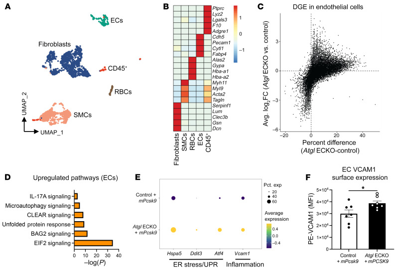
Figure 6. Endothelial deficiency of ATGL upregulates ER stress and proinflammatory gene expression in aortic ECs following a short-term atherogenic diet.
(A) Uniform manifold approximation and projection (UMAP) representation of aligned gene expression data in single cells extracted from aortas of control and Atgl ECKO mice injected with rAAV8-mPcsk9 and fed an atherogenic diet for 4 weeks. (B) Heatmap depicting gene-expression patterns of known markers of fibroblasts, SMCs, RBCs, ECs, and CD45+ immune cells. (C) Volcano plot depicting DGE patterns in the EC cluster between Atgl ECKO plus mPcsk9 compared with control plus mPcsk9 mice and expressed as log2 fold change along the y axis versus the percentage of cell expression of individual genes along the x axis. (D) Pathway enrichment of upregulated differentially expressed genes in the EC cluster between Atgl ECKO plus mPcsk9 compared with control plus mPcsk9 mice, expressed as log[–P], and analyzed by IPA. (E) Expression profiles in the EC cluster showing relative expression of the ER stress genes Hspa5, Ddit3, and Atf4 and the proinflammatory gene Vcam1 between control plus mPcsk9 and Atgl ECKO plus mPcsk9 mice. (F) Quantification of MFI of PE-VCAM1 in aortic ECs (CD31+CD45–) between control plus mPcsk9 and Atgl ECKO plus mPcsk9. n = 7 replicates/group. *P < 0.05, unpaired, 2-tailed Student’s t test.

