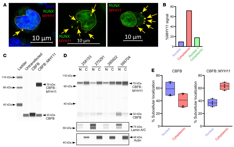
Figure 5. CBFB::MYH11 is predominantly cytoplasmic in human AML.
(A) Primary human CBFB::MYH11 AML immunofluorescence. RUNX1/2/3 is shown in green, MYH11 in red, and overlap in yellow. Yellow dashed lines outline nuclei. In a subset of images, DAPI costaining is shown in blue. Note cytoplasmic MYH11 aggregates with colocalized RUNX1 (yellow arrows) (representative images from 8 different CBFB::MYH11 patients). Scale bars: 10 μm. (B) Quantification of cells with nuclear-only MYH11, cytoplasmic-only MYH11, or both nuclear and cytoplasmic MYH11 (total of 338 cells scored). (C) K562 cells were transfected with a plasmid encoding CBFB or CBFB::MYH11, and Western blotting was performed on protein lysates using an anti-CBFB antibody. Note detection of an approximately 30 kDa band corresponding to CBFB in all lanes but increased with CBFB transfection, and detection of an approximately 79 kDa band corresponding to CBFB::MYH11 only in cells transduced with a CBFB::MYH11 plasmid. (D) ProteinSimple Jess blot on 4 human CBFB::MYH11 AML samples. Equal volumes of nuclear and cytoplasmic lysates were loaded. Anti–lamin A/C and anti-actin antibodies were used to verify nuclear and cytoplasmic purity. N, nuclear fraction; C, cytoplasmic fraction. (E) The percentage of CBFB (left panel) or CBFB::MYH11 (right panel) in the nuclear or cytoplasmic fractions of the samples shown in D. Each point represents an individual sample, bar indicates mean, box indicates 95% confidence interval, and whiskers indicate value range.

