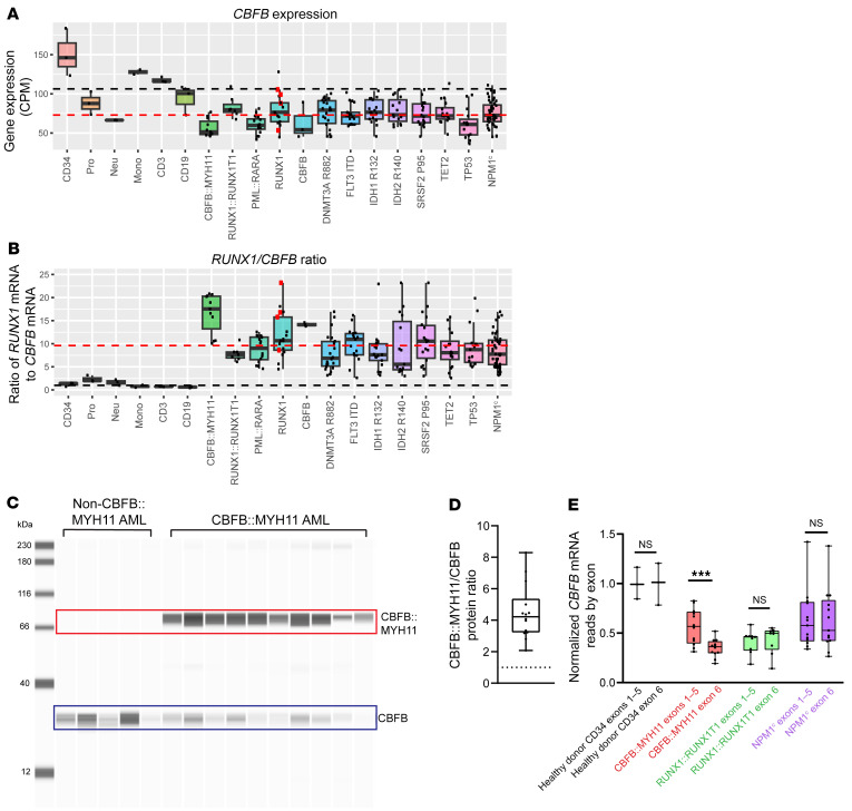
Figure 8. RUNX1/CBFB expression ratio is disrupted in human AML.
(A) TCGA LAML CBFB RNA-Seq data for the indicated healthy donor cells or AMLs. Black dashed line, mean healthy donor expression; red dashed line, mean AML expression. CBFB mRNA levels are lower in all AMLs, relative to healthy donor CD34+ cells. Each point represents an individual sample, bar indicates mean, box indicates 95% confidence interval, whiskers indicate value range. (B) Ratio of normalized, length-scaled RUNX1/CBFB mRNA expression. Black dashed line, 1:1 ratio; red dashed line, mean AML ratio. All AMLs have elevated RUNX1/CBFB ratios relative to healthy donor samples; CBFB::MYH11, RUNX1, and CBFB-mutated AMLs have the highest ratios. (C) Representative Jess blot (total of 7 experiments) of human non-CBFB::MYH11 (n = 6 patients) or CBFB::MYH11 (n = 14 patients) AML protein lysates for CBFB. Upper band (red box) indicates CBFB::MYH11; lower band (blue box) indicates WT CBFB. (D) CBFB::MYH11 to CBFB ratio in CBFB::MYH11 AMLs (n = 14 patients). Each point represents 1 patient; for patients in whom a sample was assayed more than once, point indicates mean of all assays. Dotted line, 1:1 ratio. The average CBFB::MYH11/CBFB ratio was 4.5:1, indicating that CBFB::MYH11 protein is more abundant than CBFB protein in primary human AMLs. (E) CBFB mRNA read counts from the validation cohort (Figure 7E) normalized to healthy donor CD34+ expression mean, grouped by exons 1–5 (unaffected by CBFB::MYH11 translocation) or exon 6 (lost with CBFB::MYH11 translocation). CBFB exons 1–5 expression is similar in CBFB::MYH11, RUNX1::RUNX1T1, and NPM1c AMLs, suggesting that CBFB locus transcriptional activity is unaffected by CBFB::MYH11 translocation. CBFB exon 6 reads are decreased by approximately 50% relative to exons 1–5 in CBFB::MYH11 AML, consistent with translocation-induced loss of one CBFB exon 6 allele. Paired 2-tailed t test between exons 1–5 and exon 6 reads within each group, ***q < 0.001.

