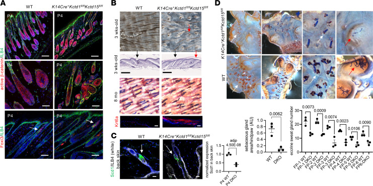
Figure 6. KCTD1/KCTD15 complexes in keratinocytes are required for the proper formation of skin appendages.
(A) Immunolabeling for active β-catenin of P4 back skin in K14Cre+Kctd1fl/flKctd15fl/fl mice and WT mice shows abnormal and shorter hair follicles in the mutant mice. Foxi3 is localized at the isthmus/infundibulum junction of hair follicles at P4 (arrows). ILB4, isolectin B4. Scale bars: 100 μm (top); 50 μm (middle and bottom). (B) Top 2 rows: Reduced hairs and abnormal hair follicles (red arrows) with flattened scale/interscale junctions (black arrows) in 3-week-old tail skin of K14Cre+Kctd1fl/flKctd15fl/fl mice. Scale bars: 250 μm. Bottom 2 rows: Abnormal hairs and reduced sebaceous glands in tail skins of 8-month-old K14Cre+Kctd1fl/flKctd15fl/fl mice, with increased Krt6a in the interfollicular epidermis. Scale bars: 10 μm. (C) Left: SCD1 immunolabeling of back skin of adult K14Cre+Kctd1fl/flKctd15fl/fl mice (DKO) and littermate controls. Scale bars, 20 μm. Right: RNA-Seq values (n = 3 per group) for Scd1 expression in P4 back skin of DKO versus control mice. Mean ± SEM; adjusted P value. (D) Oil Red O and Nile blue staining of footpad skin from adult K14Cre+Kctd1fl/flKctd15fl/fl mice shows diminished sebaceous glands (orange, white arrows) and loss of eccrine sweat glands (red arrows) compared with WT controls. Quantification of Oil Red O+ hind paw area (sebaceous glands) in 7-week-old male K14Cre+Kctd1fl/flKctd15fl/fl mice (DKO) and littermate controls. Eccrine gland duct numbers for each footpad (FP1 to FP6) of these mice are shown. Mean ± SEM; P values (2-tailed t test; n = 3 mice per group).

