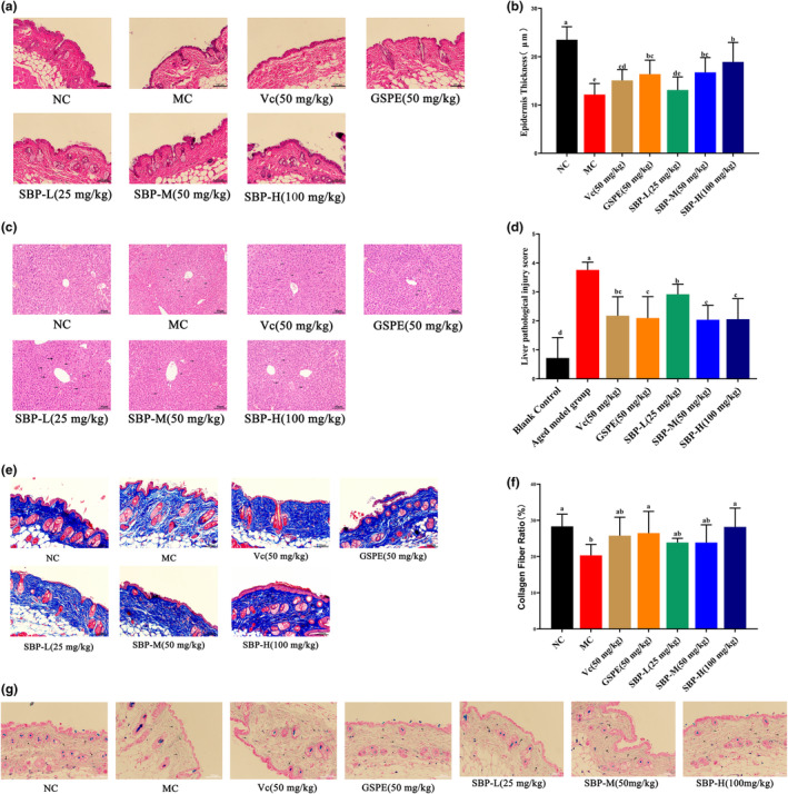
FIGURE 4.

Histopathological observation of skin and liver and the effect of SBP on collagen and elastic fibers in the skin of aging mice. (a) The results of H&E staining on mice skin (200×, scale bar 100 μm). (b) Quantification of epidermal layer thickness based on skin H&E staining results. (c) The results of H&E staining on mice liver tissue (200×, scale bar 40 μm). (d) Liver pathological injury score. (e) The results of Masson staining on mice skin (200×, scale bar 100 μm). (f) Quantification of the proportion of collagen fibers based on skin Masson staining. (g) The results of Victoria blue staining on mice skin (200×, scale bar 100 μm). a‐fMean values within the same row not sharing a common superscript letter are significantly different (p < .05) by Duncan's multiple range test.
