
Figure 3: UroA impairs iAs3+ induced ROS generation and protects against oxidative stress.
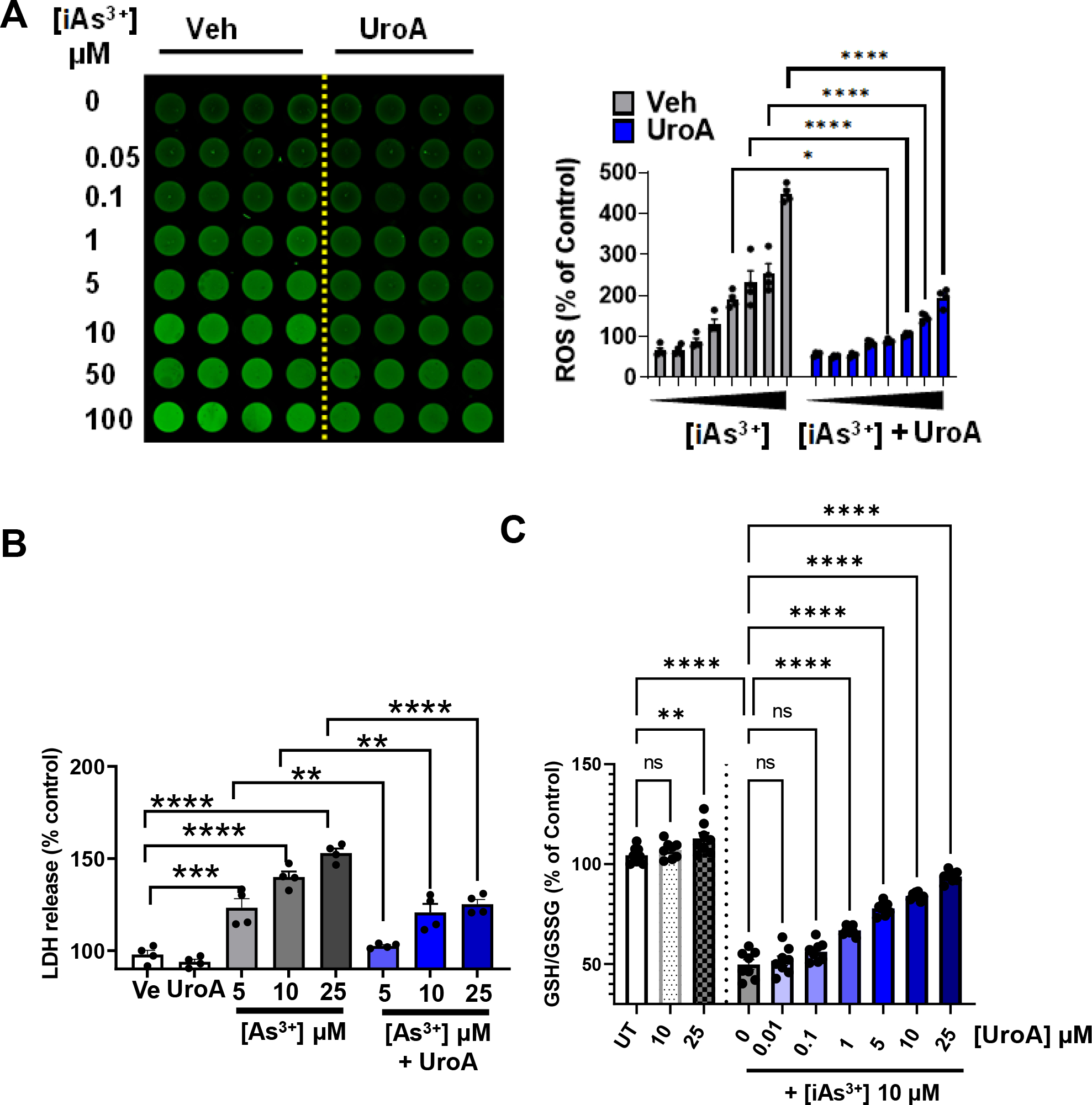
(A) T84 cells were treated with iAs+3 (0, 0.05, 0.1, 1, 5, 10, 50, 100 μM) in presence of vehicle (DMSO-0.01%) or UroA (25 μM) for 12 h. Representative fluorescence image and bar graph are showing ROS generation as green fluorescence from DCFDA stained T84 cells. (B) LDH release after 24 h incubation of T84 cells with iAs+3 (5, 10, 20 μM) in presence of vehicle (DMSO-0.01%) or UroA (25 μM) (C). T-84 cells were treated with UroA (10, 25 μM). T84 cells were treated with iAs+3 (10 μM) and different concentration of UroA (0, 0.01, 0.1, 1, 5, 10, 25 μM) for 24 h. The levels of GSH and GSSG were measured and the GSH/GSSG ratio was calculated. Untreated (UT) cells were used as control (100%). Results are representative of three independent experiments. Statistics performed using 2way ANOVA in GraphPad Prism software. *p < 0.05, **p < 0.01, ***p < 0.001, ****p < 0.0001. Error bar, mean ± SEM (n=4–8)