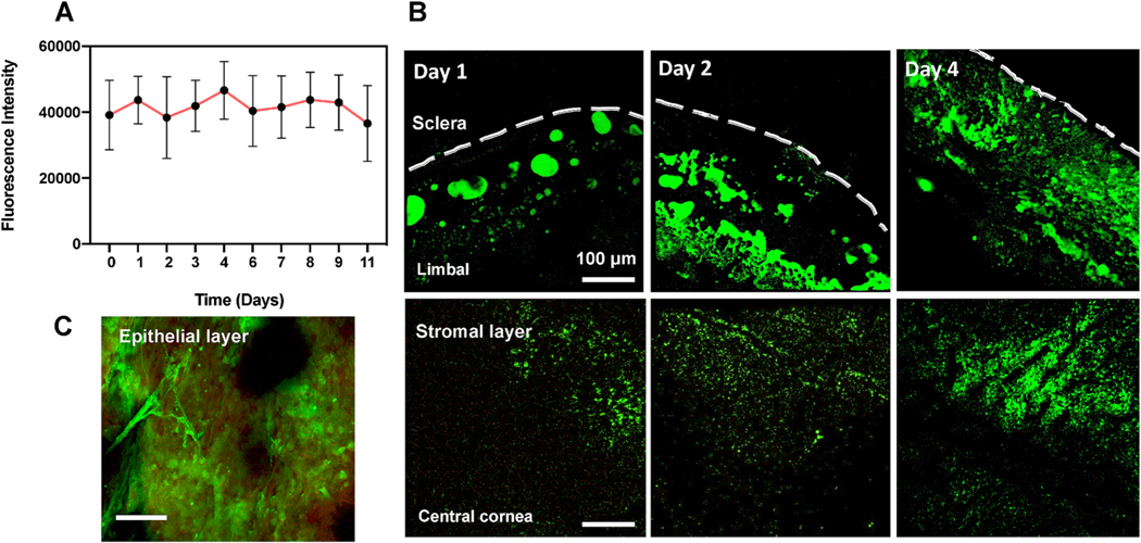
Fig. 4. Fluorescence data showing s-HA hydrogel application to wounded cornea.
(A) Fluorescence intensity of s-HA-FITC after 10 days in PBS. (B) Confocal images show s-HA-FITC in the limbal and central corneas at days 1, 2 and 4 in the stromal layer after corneal injury. Scale bar: 100 μm. (C) HA-FTIC (green) adsorbed into the epithelial layer (red) near limbal area.

