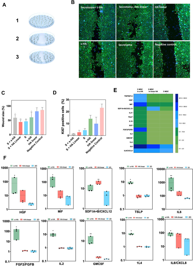
Fig. 5. s-HA hydrogel modulates in vitro CEC wound healing and c-MSC secretome profile.
(A) Schematic of in vitro wound healing model. (B) Ki67 (magenta) and F-actin (green) expression in CECs treated with (1) secretome from c-MSCs encapsulated in s-HA hydrogel, (2) c-MSCs encapsulated in linear HA (3) linear HA, (4) s-HA hydrogel, (5) secretome alone and (6) complete medium (negative control) 72 h after wound (scale bar: 100 pixel). (C) Quantification of the wound size (%) 72 h after wound (n = 3). Wound size significantly decreased for the CECs treated with secretome from c-MSCs encapsulated in s-HA hydrogel and with secretome from c-MSC encapsulated in linear HA hydrogel compared to negative control. CEC wound size also decreased by treating the cells with s-HA hydrogel compared to negative control. Ordinary one-way ANOVA (p < 0.005) was used to detect statistical differences followed by uncorrected Fisher LSD (****p < 0.0001, ***p = 0.0001, **p <0.05 vs negative control) (D) Quantification of Ki67 positive cells 72 h after wound showing expression of ki67 positive cells for CEC treated with (1) secretome from c-MSCs encapsulated in s-HA hydrogel, (2) c-MSCs encapsulated in linear HA (3) linear HA, (4) s-HA hydrogel, (5) secretome alone and (6) complete medium. (E) Heat map showing the different protein expression profiles for c-MSCs encapsulated in s-HA and linear HA viscoelastic solution and 2D condition. The different colors represent relative Mean fluorescence Intensity (MFI) normalized to 2D condition (F) c-MSCs encapsulated within s-HA and linear HA significantly increase the expression of growth factors and inflammatory and pro-inflammatory cytokines and chemokines compared to c-MSCs in TCP (2D condition). Kruskal–Wallis H test followed by uncorrected Dunn’s test was used to detect statistically significant differences between groups.

