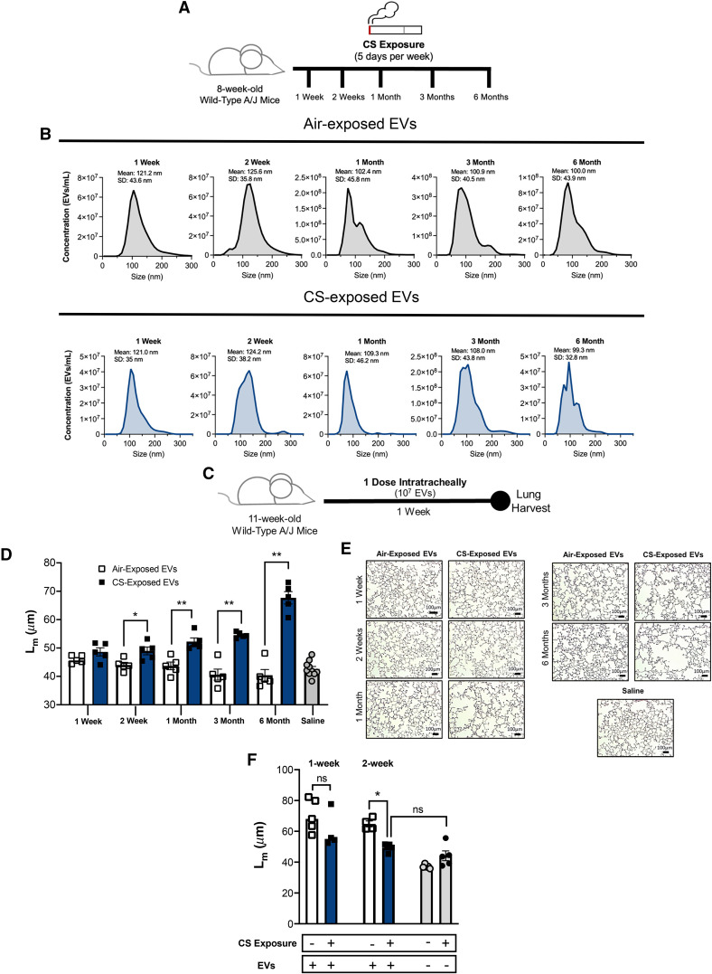
Figure 1.
Transfer of cigarette smoking (CS)–exposed extracellular vesicles (EVs) induces alveolar damage in naive recipients. (A) Airway EVs from mice receiving cigarette smoke for 1 week, 2 weeks, 1 month, 3 months, or 6 months were harvested, purified, counted. (B) Their size distribution was determined using Nanosight NS300. Histograms represent EV samples pooled from five individual mice. Data are representative of two or more independent exposure experiments. (C) EVs were transferred to naive A/J mice intratracheally. Mice were treated with one dose (107 EVs), and their lung damage was quantified 1 week posttreatment (n = 5 per group). (D) Cumulative mean linear intercept (Lm) scoring as a means of quantifying alveolar enlargement (n = 5 per group). (E) Representative images (hematoxylin and eosin) 1 week after EV transfer. Scale bars, 100 μm. (F) A/J mice underwent smoke or air exposure for a 1-week or 2-week period. After exposure, mice were treated with one dose of 3-month, smoke-activated EVs (107 EVs), and their lung damage was quantified 1 week posttreatment (n = 4 or 5 per group). The quantified results are expressed in terms of mean ± SEM. For all comparisons, statistical significance was determined by Mann-Whitney test. *P < 0.05 and **P < 0.01. ns = not significant.

