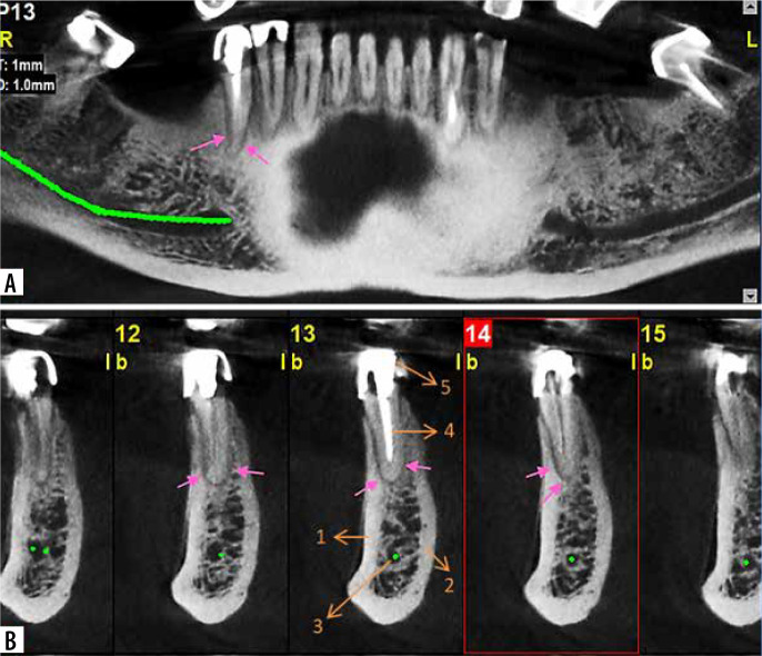
Figure 10.
Panoramic-like (A) and cross-sectional (B) cone-beam computed tomography sections that demonstrate radiolucency around the apex of the right second mandibular premolar (pink arrows), presenting apical periodontitis. 1) Buccal cortical plate, 2) lingual cortical plate, 3) mandibular canal, 4) root, 5) crown

