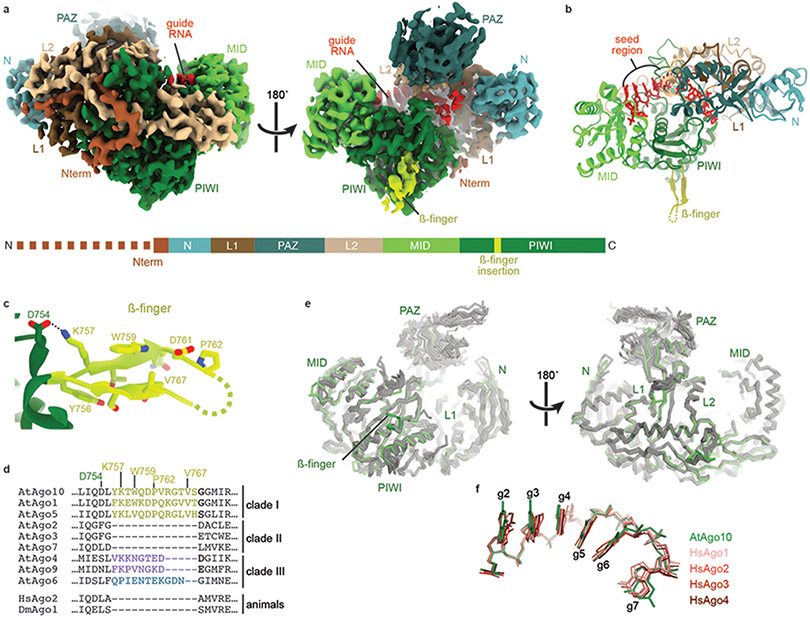
Fig. 1. Structure of the AtAgo10-guide RNA complex without target RNA.
a. Cryo-EM reconstruction with individual domains segmented and colored as in the schematic (lower panel). The dashed line indicates unstructured N-terminal residues. Guide RNA density colored red. b. Cartoon representation of the AtAgo10-guide RNA atomic model. c. Close-up of the ß-finger structure. d. Sequence alignment near the ß-finger insertion of AGOs from Arabidopsis, human, and Drosophila. Plant AGO clades indicated. e. Cα backbone superposition of AtAgo10-guide structure (green) and human AGO-guide (silver) crystal structures. f. Superposition of guide nucleotides (shown as sticks) from the seed regions of AtAgo10 (green) and representatives of the four human AGOs (shades of red).

