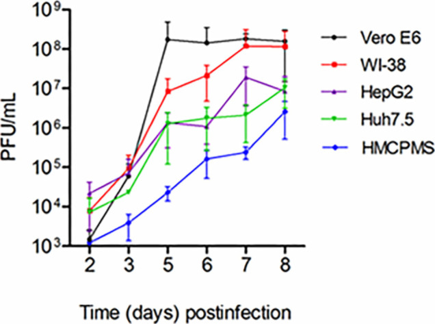
Figure 1.

SARS-CoV-2 virus replication. Kidney (Vero E6), lung (WI-38), liver (HepG2, Huh7.5) and human primary hepatocytes HMCPMS were infected with SARS-CoV-2 at MOI of 0.1. About 70%-80% confluent cells were incubated with the virus for 1 hour. After incubation, wells were washed with PBS (3X) and replaced with fresh growth media. Postinfection media was collected from each well on the days indicated and analyzed by plaque assay for viral growth kinetics. (PFU/mL =plaque-forming units/mL) Error bars represent standard deviation among cell samples, n=3.
