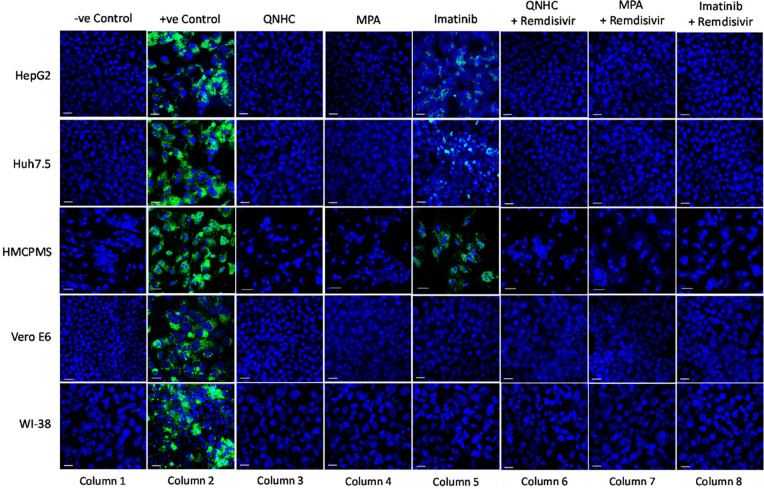
Figure 5.
SARS-CoV-2 infection inhibition on cell lines preincubated with antiviral and viral inhibitor commixture. Cell lines, 70% to 80% confluent, were preincubated with 5µM quinacrine dihydrochloride (QNHC), 4µM mycophenolic acid (MPA), and 10µM imatinib and a commixture of QNHC, 5µM +5 µM remdesivir, MPA, 4µM + 5µM remdesivir, and imatinib, 10 µM + 5µM remdesivir for 24 hours in a 12.5 mm polyester membrane transwell plate. Following incubation, the wells were infected with 0.1 MOI of SARS-CoV-2 virus for 1 hour, washed with PBS (3X), and replaced with complete growth media. Infected cells were further incubated for 3 days before being stained for SARS-CoV-2 spike proteins. Virus only was used as positive control and no virus as negative control. Fluorescent signal was captured using Zeiss LSM710 LIVE Duo confocal microscope. Bar indicates 20µm. n=3

