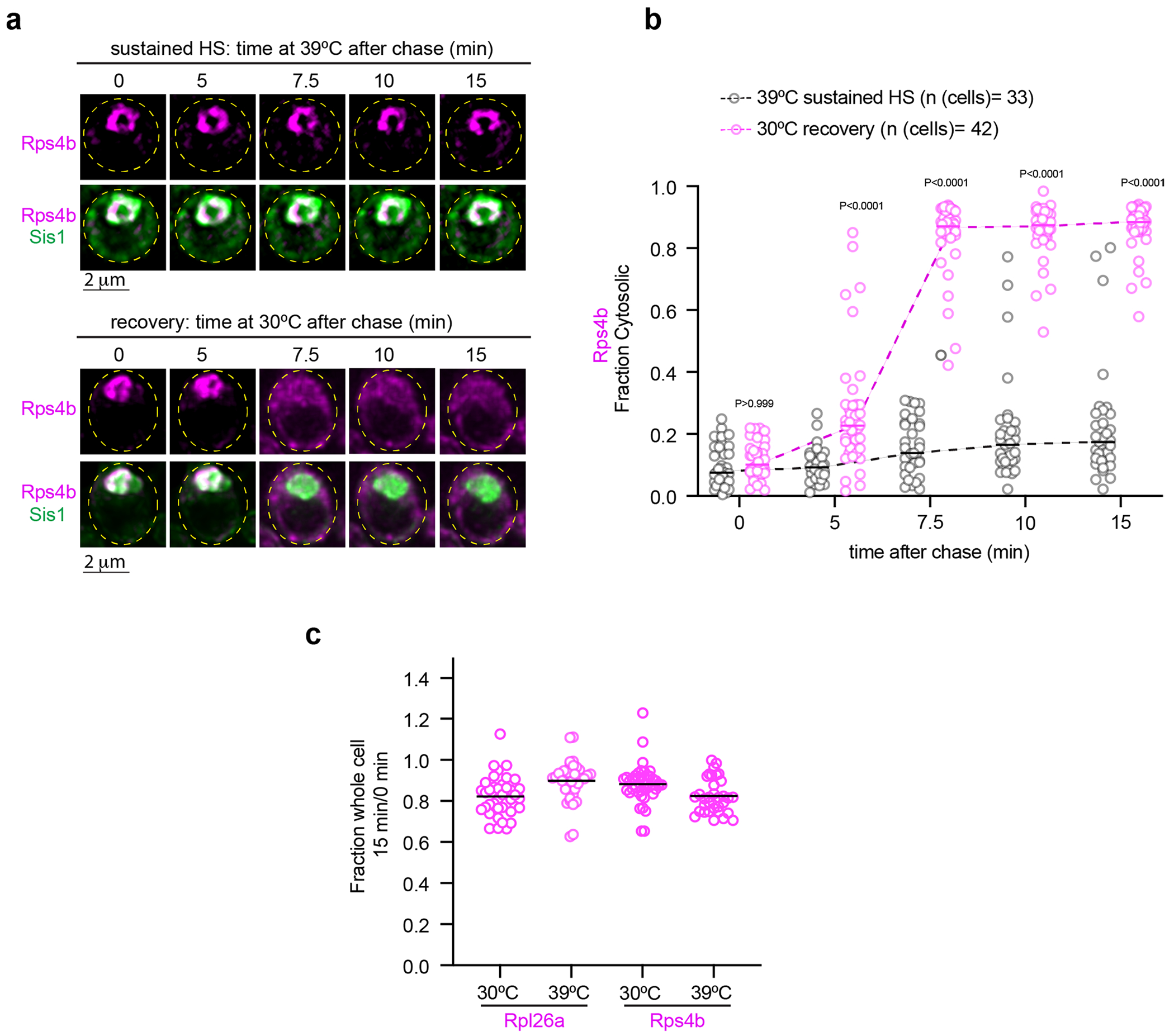
Extended Data Fig. 8 |. oRPs in condensates are not degraded and are transported to the cytosol upon recovery.

(a) Live cell time lapse imaging of the spatial distribution of Rps4b (magenta) and Sis1-mVenus (green) during sustained heat shock and recovery. (b) Quantification of the fraction of cytosolic Rps4b signal under sustained HS or recovery. Statistical significance was established using the Brown-Forsythe and Welch one-way ANOVA test, followed by Dunnett T3 multiple comparison analyses. n = number of cells pooled from 3 biologically independent replicates. (c) Fraction of total pulse labeled Rpl26a or Rps4b remaining after chase for 15 minutes at indicated temperature. n=number of cells pooled from 3 biologically independent replicates.
