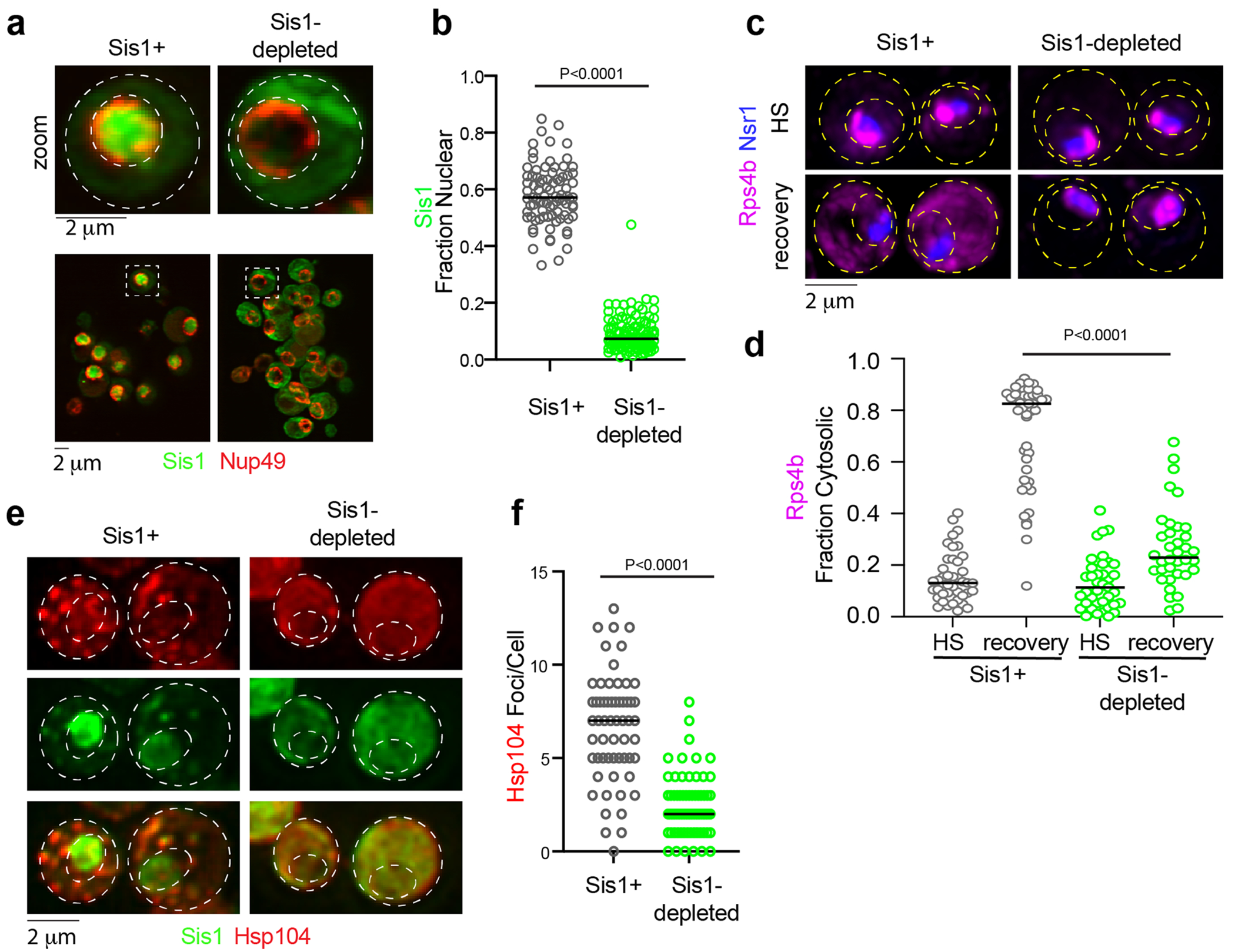
Extended Data Fig. 9 |. oRP condensate reversibility depends upon Sis1 availability.

(a) imaging of Sis1-mVenus (green) and Nup49-mScarlet-I (red) following Sis1 depletion or not. (b) Quantification of fraction of nuclear Sis1 upon Sis1 depletion or not. P values were calculated with unpaired two-tailed Welch’s t-test. n denotes number of cells as obtained from 4 independent experiment. n = number of cells pooled from 4 biologically independent replicates. (c) LLS imaging of Hsp104-mKate2 during heat shock (39 °C, 15 mins) pre-depleted for Sis1 (green) or not. (d) Quantification of Hsp104-mKate2 foci per cell for (c). P values were calculated with unpaired two-tailed Welch’s t-test. n denotes number of cells from 3 independent experiments. n = number of cells pooled from 3 biologically independent replicates. (e) LLS live cell imaging of Rps4b (magenta) and the nucleolar marker Nsr1 (blue) during heat shock and recovery in the absence or presence of Sis1 depletion. (f) Quantification of the fraction of cytosolic Rps4b under sustained HS or recovery in the absence or presence of Sis1 depletion. P values were calculated with unpaired two-tailed Welch’s t-test. n denotes number of cells obtained from 3 independent experiment. n = number of cells pooled from 3 biologically independent replicates.
