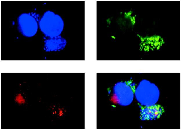
FIG. 3.
Confocal laser scanning microscopy of HeLa cells infected with wild-type EPEC. Bacterial and cellular nucleic acid was labeled with DAPI (blue). Actin was labeled with fluorescein isothiocyanate-phalloidin (green). EspB was labeled with affinity-purified EspB antiserum and detected with a secondary antibody against immunoglobulin G conjugated to lissamine rhodamine (red). Areas of colocalization of EspB and actin appear yellow.

