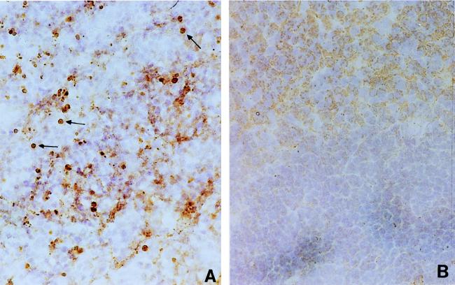
FIG. 3.
Apoptosis in spleen cells in H. capsulatum-infected mice. Spleen cryosections of H. capsulatum-infected (2 to 3 weeks postinfection) (A) and normal (B) mice were stained with TUNEL reagents, and DAB substrate was used for color development. The arrows point to brown apoptotic nuclei. Magnification, ×200.

