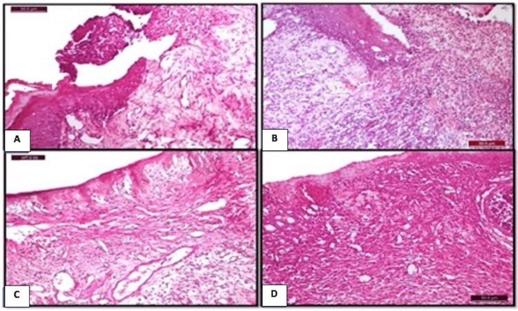
Figure 3. Photomicrograph of the different groups at day 7 post-ulceration induction.
A) Control group: showing epithelial proliferation at the lateral side of the ulcer site (H&E x400). B) CPC-treated group: showing coagulative mass mixed with PMNLs protruded from the wound site and epithelium proliferation at the lateral border (H&E x100). C) NS-treated group: showing the formation of thin epithelial tissue covering the ulcerative site and the subepithelial connective tissues composed of well-organized granulation tissues with abundant vascularity (H&E x200). E) WGO-treated group: showing proliferated epithelial tissue covering the ulcerative site, with the formation of heavily vascularized granulation tissues in the subepithelial connective tissues (H&E x100).
CPC: cetylpyridinium chloride; PMNLs: polymorphonuclear leukocytes; NS: Nigella sativa; WGO: wheat germ oil; H&E: hematoxylin and eosin

