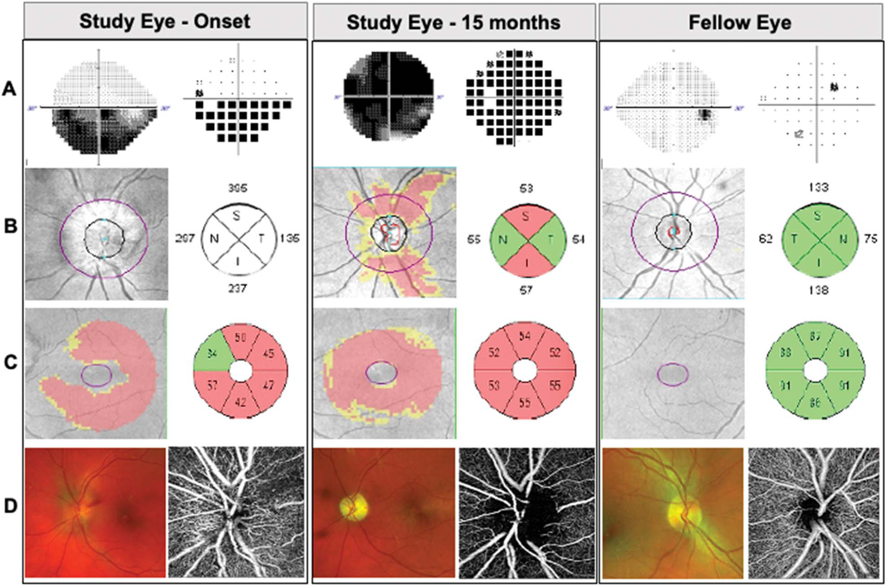
FIG. 1.

Representative patient with high-altitude NAION in left eye. Images demonstrating study eye at onset and, at 15-month follow-up, and fellow eye. A. Static perimetry revealed inferior altitudinal visual field defect that worsened over time. Gray scale (left) and pattern deviation (right). B. Spectral-domain optical coherence tomography (OCT) and peripapillary retinal nerve fiber layer thickness deviation map and quantification by quadrants (in microns). C. OCT macular ganglion cell complex thickness deviation map and quantification by sectors (in microns). D. Scanning laser ophthalmoscopy images of the optic disc (Optos, left) and OCT angiography of the optic disc (Zeiss AngioPlex, right).
