
Fig. 5.
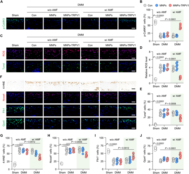
MNPs-TRPV1 protects chondrocytes from ferroptosis under AMF stimulation. (A and B) Representative images (A) and corresponding quantification (B) of the immunofluorescence staining of p-CaMKII in the mouse articular cartilage treated with MNPs or MNPs-TRPV1 with or without AMF exposure. (C to E) Measurement of ROS and Tunel+ chondrocytes (C), and their quantification (D and E) in the articular cartilage with the indicated treatment. (F to J) Representative images (F) of immunohistochemical or immunofluorescence staining and their quantification analysis of 4-HNE (G), Ncoa4 (H), Fth1 (I), and Gpx4 (J) in the mice cartilage treated with MNPs or MNPs-TRPV1 with or without AMF exposure. w/o, without; w/, with. Scale bars, 50 μm. One-way ANOVA with Tukey’s post hoc test. Data are shown as mean ± SD.