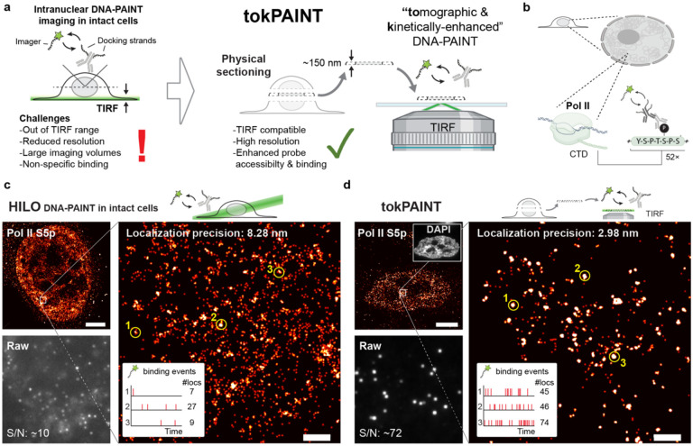
Figure 1 |. tokPAINT enables TIRF-based DNA-PAINT imaging of intranuclear targets.
a tokPAINT schematic. Ultrathin cryosectioning enables nuclear DNA-PAINT imaging under TIRF conditions. b Immunolabeling of Pol II CTD Serine-5 phosphorylation (S5p) for DNA-PAINT imaging via docking strand-conjugated secondary antibodies. c Top leZ: HILO DNA-PAINT image of Pol II S5p within intact HeLa cell. Bottom left: frame from HILO DNA-PAINT raw data acquisition including signal-to-noise ratio (S/N). Right: magnified region as indicated by white square. Time traces of imager binding and number of localizations are shown for three regions indicated by yellow circles. d Top left: tokPAINT image of Pol II S5p. The inset shows the same cell imaged in the DAPI channel. Bottom left: frame from tokPAINT raw data acquisition including S/N ratio. Right: magnified region as indicated by white square. Time traces of imager binding and number of localizations are shown for three regions indicated by yellow circles. Scale bars, 5 μm in (c,d), 400 nm in zoom-ins.

